
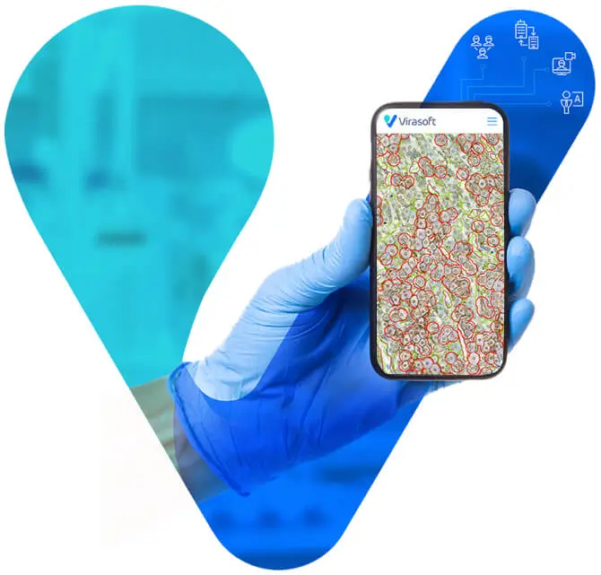
Explore Virasofts advanced academic platforms designed for the future of pathology.
Virasoft’s academic solutions are consisted of:
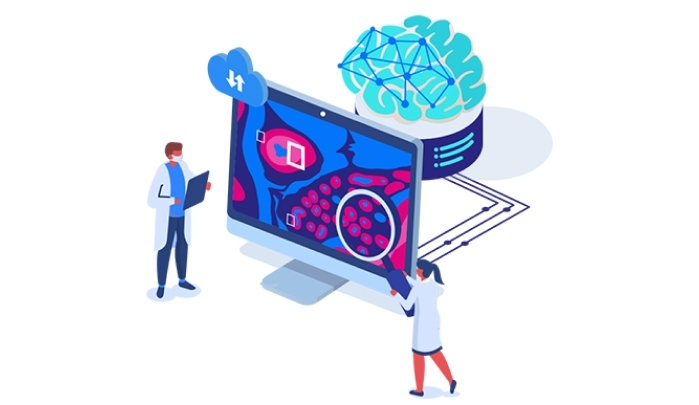
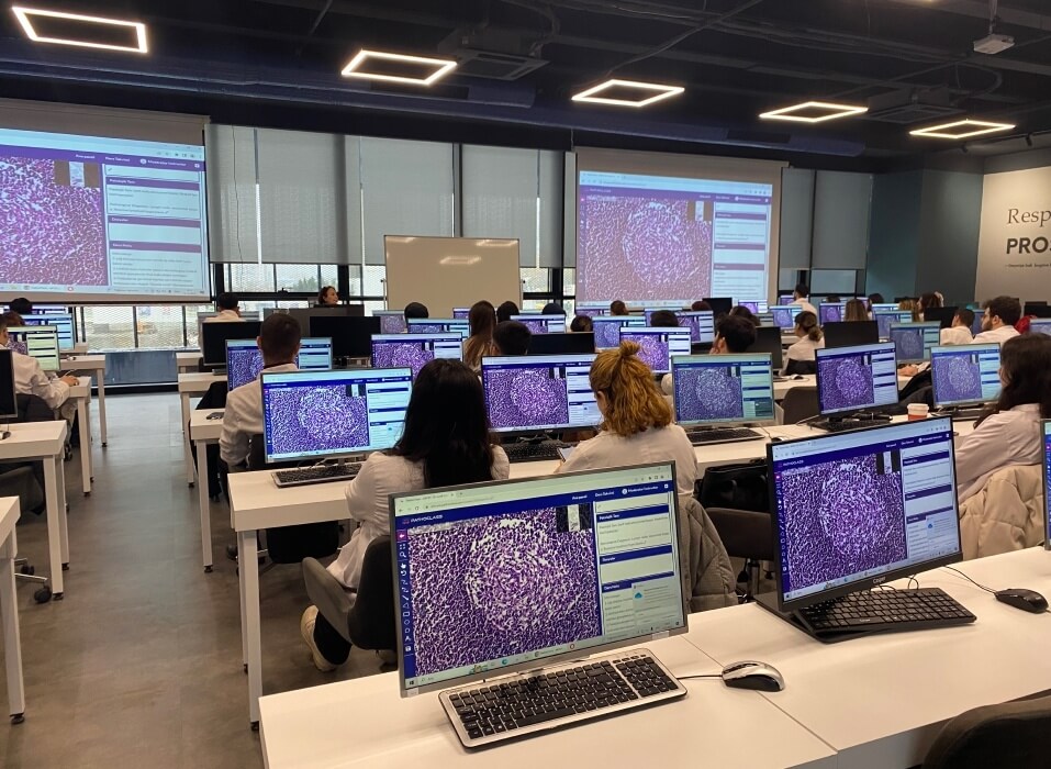
Our academic platforms bring instructors and students together with scanned whole slide images (WSIs), assignments and lectures.


Virasoft Solution: VIRAPIS
Virasoft Solution: VIRAPIS
Virasoft Solution: VIRAPIS
Virasoft Solution: VIRAPIS
Virasoft Solution: VIRAPIS
Virasoft Solution: VIRAPIS
Virasoft Solution: VIRASIGHT
Virasoft Solution: VIRAPIS
Virasoft Solution: VIRAPIS & VIRASIGHT
cases reported
in 2025
algorithms used
in 2025
cases consulted
in 2025
students attended
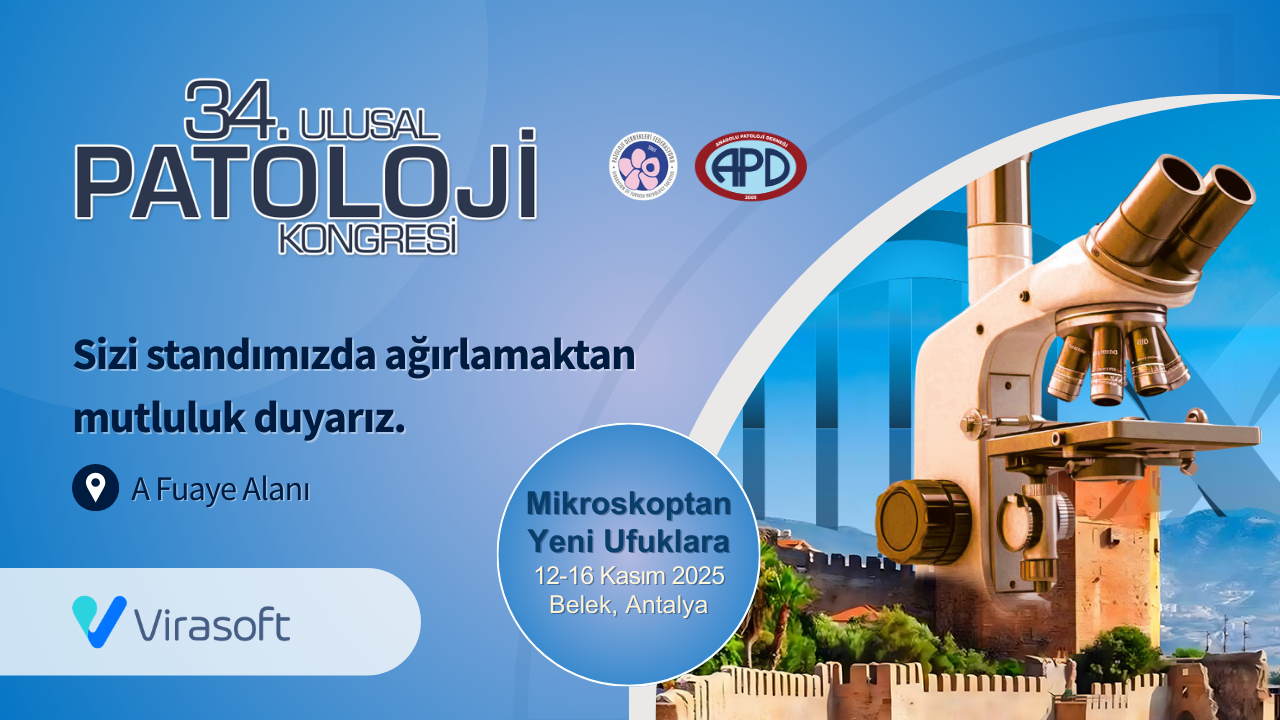
Virasoft presented its innovative digital pathology solutions at the 34th National Pathology Congress and attracted strong interest with its satellite symposium, “Introduction to Digital Pathology in 10 Questions.”
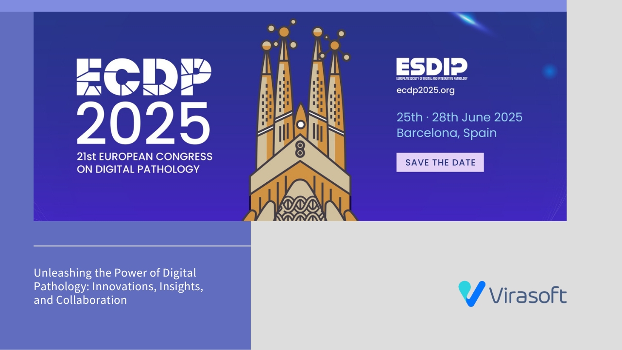
Virasoft presented 3 poster studies at ECDP 2025, highlighting its latest AI-driven innovations in pathology. From invasive area detection in breast cancer to NLP-based data extraction and digital workflow transformation, these studies marked a step beyond image processing and redefined the potential of digital pathology.

Virasoft has successfully implemented its digital and AI-driven solutions at Pathoteam Diagnostic, enhancing diagnostic accuracy, workflow efficiency, and collaboration. This partnership marks a significant step in advancing pathology practices with cutting-edge technology.
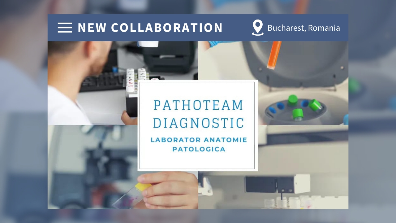
Virasoft and Pathoteam Diagnostic join forces to set new standards in pathology with advanced AI solutions.
This collaboration represents a significant milestone in Virasoft’s mission to provide innovative AI-powered pathology solutions to laboratories around the globe.
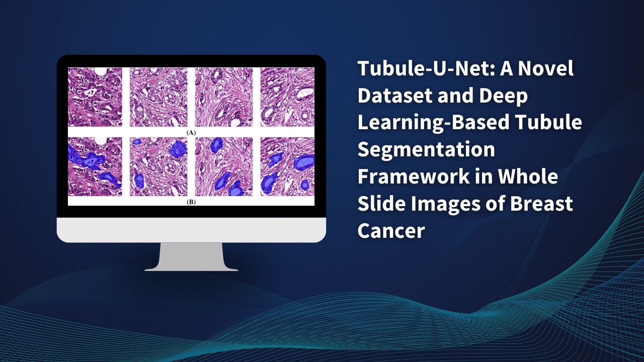
Virasofts research article, "Tubule-U-Net: A novel dataset and deep learning-based tubule segmentation framework in whole slide images of breast cancer," published in Scientific Reports (Nature, 2023), ranked among the top 100 most downloaded cancer research papers of the year.