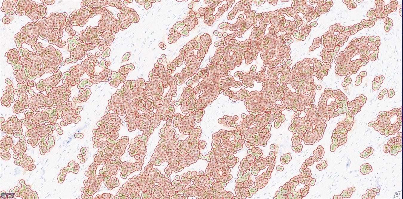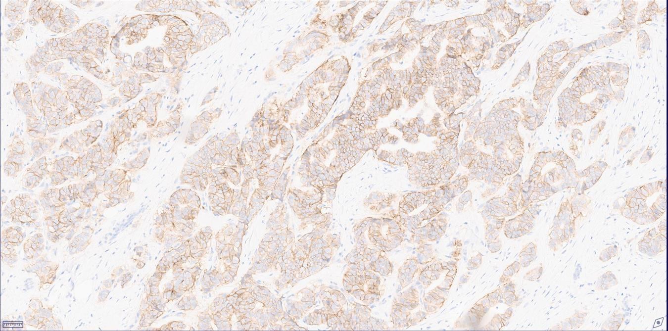
One of the methods to determine HER2 protein expression is immunohistochemistry (IHC) [1]. IHC stained tissues are evaluated according to American Society of Clinical Oncology/College of American Pathologists (ASCO/CAP) HER2 guidelines. According to guidelines, the scoring method for HER2 IHC is semiquantitative and is based on 4 classes (0/ 1+, 2+, 3+). Manual HER2 evaluation of IHC stained slides is susceptible to inter-observer variability and involves an error-prone.Virasoft offers reproducible, automatic, and objective analysis and interpretation of selected regions of interest (ROI) as well as whole slide images (WSI) that aimed to aid pathologists for more time-efficient cell counting process. By IHC, only a score of 3+ is reported as positive for HER2 amplification. All 2+ equivocal cases have to undergo subsequent testing by FISH [2].We also developed HER2 algorithm for cost-efficient diagnostic process for medical centers.
Keywords: Membrane Segmentation, HER2 Assessment, Breast Cancer, Gastric Cancer Digital Pathology, ASCO/CAP
Methods:
Automatic HER2 scoring is the ability to give consistent results on similar slides in a short time compared with the manual scoring performed by pathologists. Virasoft HER2 Analyzer is developed based on preprocessing, thresholding and segmentation techniques to score the whole slide images. The technique is comprised of three steps. In the first step, a superpixel-based support vector machine (SVM) feature learning classifier is proposed to classify epithelial and stromal regions from WSI. In the second stage, on classified epithelial regions, a convolutional neural network (CNN) based segmentation method is applied to segment membrane regions. Finally, divided tiles are merged and the overall score of each slide is evaluated.
Quantitative output variables:
HER2 IHC Score
Strong Cell (+3) Count and Percentage
Medium Cell (+2) Count and Percentage
Weak Cell (+1) Count and Percentage
Incomplete Cell (0) Count
Workflows:
View the HER2 whole slide digital image with ViraPath.
Outline tumor either manually or automatically using Virapath Tissue Segmentation algorithm.
Select HER2 analysis and calibrate the parameters.
Run the analysis.
References
[1] Loibl, S., & Gianni, L. (2017). HER2-positive breast cancer. The Lancet, 389(10087), 2415–2429.
[2] Antonio C Wolff, M Elizabeth H Hammond, ... Gail H Vance, Giuseppe Viale, Daniel F Hayes, ASCO; CAP, Recommendations for human epidermal growth factor receptor 2 testing in breast cancer: American Society of Clinical Oncology/College of American Pathologists clinical practice guideline update, Arch Pathol Lab Med. 2014 Feb;138(2):241-56.
[3] Automated segmentation of cell membranes to evaluate HER2 status in whole slide images using a modified deep learning network, Fariba Damband Khameneh, Salar Razavi, Mustafa Kamasak, Computers in Biology and Medicine, 2019.
